The Brain
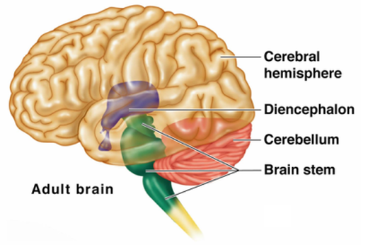
The central nervous system (CNS) consists of the brain and the spinal cord. The brain is the largest and most complex part of the nervous system. It includes the two cerebral hemispheres , the diencephalon . the brain-stem (which attaches the brain to the spinal cord), and the cerebellum. The brain includes about one hundred billion multipolar neurons and countless branches of the axons by which these neurons communicate with each other and with neurons elsewhere in the nervous system. The brain-stem connects the brain and spinal cord and allows two-way communication between them. The spinal cord, in turn, provides two-way communication between the CNS and the peripheral nervous system (PNS). Bones, membranes, and fluid surround the organs of the CNS. More specifically, the brain lies within the cranial cavity of the skull, whereas the spinal cord occupies the vertebral canal within the vertebral column. Beneath these bony coverings, membranes called meninges , located between the bone and the soft tissues of the nervous system, protect the brain and spinal cord (Shier 2007). The brain contains nerve centers associated with sensory functions and is responsible for sensations and perceptions. It issues motor commands to skeletal muscles and carries on higher mental functions, such as memory and reasoning. It also contains centers that coordinate muscular movements, as well as centers and nerve pathways that regulate visceral activities. In addition to overseeing the function of the entire body, the brain also provides characteristics such as personality.
4 Regions of the Brain:
4 Regions of the Brain:
- Cerebral Hemispheres (cerebrum)
- Diencephalon
- Brain Stem
- Cerebellum
Essentials of Human Anatomy and Physiology, 2009
Cerebrum
The cerebrum , otherwise known as the telencephalon, makes up about 80 percent of the brain's weight. It lies above the diencephalon. The cerebrum's outer layer, the cerebral cortex, is made entirely of gray matter. White matter makes up the inner portion of the cerebrum. The tissue of the cerebral cortex is about 0.08 to 0.16 inch (2 to 4 millimeters) thick. The cerebral cortex is folded extensively. The folds are called convolutions or gyri, and the shallow grooves between the folds are sulci. Deeper grooves, which are less numerous, are called fissures. The folds greatly increase the surface area of the cerebral cortex--it would have a surface area of about 5 square feet (1.5 square meters) if spread out--and thus the total number of nerve cell bodies it contains. Located deep within the white matter of the cerebrum just above the diencephalon are two paired masses of gray matter known as basal ganglia (Gale 2007). They are important in coordinating subconscious skeletal muscular movement such as swinging the arms while walking. A deep fissure separates the cerebrum into a left and right hemisphere or half. The corpus callosum, a bundle of more than 200 million neurons, connects the two cerebral hemispheres and carries vast amounts of information between them--an estimated 4 billion nerve impulses per second. By studying patients whose corpora callosa had been destroyed, scientists have learned that some differences exist between the left and right hemispheres. The left side of the brain functions mainly in speech, logic, writing, and arithmetic. The right side of the brain, on the other hand, is more concerned with imagination, art, symbols, and spatial relations. These distinctions should not be pushed too far, however, as studies of patients with brain injuries indicate that various functions associated with one side of the brain can often be recovered by the other side (2007).
Right and Left Side of the Brain:
Lobes of the Cerebrum:
Frontal Lobe:
The frontal lobe forms the anterior portion of each cerebral hemisphere. It is bordered posteriorly by a central sulcus (fissure of Rolando), which passes out from the longitudinal fissure at a right angle, and inferiorly by a lateral sulcus (fissure of Sylvius), which exits the under surface of the brain along its sides (Shier 2007). Controls:
Parietal Lobe:
The parietal lobe is posterior to the frontal lobe and is separated from it by the central sulcus. Controls:
Occipital Lobe:
The occipital lobe forms the posterior portion of each cerebral hemisphere and is separated from the cerebellum by a shelf-like extension of dura mater called the tentorium cerebelli (2007). The occipital lobe and the parietal and temporal lobes have no distinct boundary. Controls:
Temporal Lobe:
The temporal lobe lies inferior to the frontal and parietal lobes and is separated from them by the lateral sulcus. Controls:
- Cerebral Hemispheres (Cerebrum)
- Paired (left and right) superior parts of the brain
- Included more than half of the brain mass
- The surface is made of ridges (gyri) and grooves (sulci)
Right and Left Side of the Brain:
- Each hemisphere controls the opposite side
- Right side controls: creativity, spatial ability, artistic and musical skills
- Left side controls: speech, comprehension. arithmetic, and writing
- We all use both side but some sides may be more dominant than the other for most people
Lobes of the Cerebrum:
- Fissures (deep grooves) divide the cerebrum into lobes
- Surface lobes include:
- Frontal Lobe
- Parietal Lobe
- Occipital Lobe
- Temporal Lobe
Frontal Lobe:
The frontal lobe forms the anterior portion of each cerebral hemisphere. It is bordered posteriorly by a central sulcus (fissure of Rolando), which passes out from the longitudinal fissure at a right angle, and inferiorly by a lateral sulcus (fissure of Sylvius), which exits the under surface of the brain along its sides (Shier 2007). Controls:
- Personality, behavior, emotions
- Judgement, planning, problem solving
- Speech: speaking and writing (Broca's area)
- Body movement (motor strip)
- Intelligence, concentration, and self awareness
Parietal Lobe:
The parietal lobe is posterior to the frontal lobe and is separated from it by the central sulcus. Controls:
- Interprets language, words
- Sense of touch, pain, and temperature (sensory strip)
- Interprets signals from vision, hearing, motor, sensory, and memory
- Spatial and visual perception
Occipital Lobe:
The occipital lobe forms the posterior portion of each cerebral hemisphere and is separated from the cerebellum by a shelf-like extension of dura mater called the tentorium cerebelli (2007). The occipital lobe and the parietal and temporal lobes have no distinct boundary. Controls:
- Interprets vision (color, light, movement)
Temporal Lobe:
The temporal lobe lies inferior to the frontal and parietal lobes and is separated from them by the lateral sulcus. Controls:
- Understanding language (Wernicke's area)
- Memory
- Hearing
- Sequencing and organizing
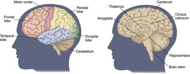
The anatomy of the human brain, exterior view showing the lobes (left) and the interior sections (right).
http://ic.galegroup.com/ic/scic/ImagesDetailsPage/ImagesDetailsWindow?total=2&query=BS lobes&prodId=SCIC&windowstate=normal&mode=view&limiter=AC y&displayGroupName=Images&currPage=1&sortBy=relevance%2Cdescending&action=e&catId=&view=docDisplay&documentId=GALE%7CCV2210038593&userGroupName=22503&jsid=2bea0882d5a387ce3394a0735e5d8ab6
Layers of the Cerebrum:
- Gray matter -- outer layer in the cerebral cortex composed mostly of neuron cell bodies
- White matter -- fiber tracts deep to the gray matter
- Corpus callosum connects right and left hemispheres
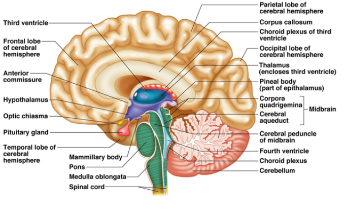
Diencephalon
The diencephalon is the part of the fore-brain that lies above the brain stem. It includes the thalamus and hypothalamus. The thalamus is an important relay station for sensory information coming to the cerebral cortex from other parts of the brain. The thalamus also interprets sensations of pain, pressure, temperature, and touch, and is concerned with some of our emotions and memory. It receives information from the outside environment in the form of sounds, smells, and tastes (Gale 2010). The hypothalamus performs numerous important functions. These include the control of the autonomic nervous system. The hypothalamus controls normal body temperature and helps regulate the endocrine system, which produces hormones or chemical messengers that regulate body functions. It informs the body of hunger, fullness, or thirst. It helps regulate sleep and wakefulness and is involved in the emotions of anger and aggression.
Thalamus:
The thalamus is a selective gateway for sensory impulses ascending from other parts of the nervous system to the cerebral cortex. It receives all sensory impulses (except those associated with the sense of smell) and channels them to appropriate regions of the cortex for interpretation. In addition, all regions of the cerebral cortex can communicate with the thalamus by means of descending fibers. The thalamus transmits sensory information by synchronizing action potentials (Shier 2007).
Hypothalamus:
The hypothalamus maintains homeostasis by regulating a variety of visceral activities and by linking the nervous and endocrine systems.
Epithalamus:
In terms of function, the epithalamus is responsible for connecting the lymbic system to the rest of the brain, as well as regulating hormones secreted by the pineal gland. Looking at a side view of the human brain, the epithalamus is located just above the medulla oblongata and the pituitary gland. Following the path of the spinal cord into the skull, the cord transitions into the brain via the medulla oblongata, ending in the lower section of the middle brain. Just above and a few centimeters forward, rests the pituitary gland on a horizontal axis with the eyes and ears. Extending over both the medulla oblongata and the pituitary gland is the epithalamus and its associated components (2010).
- Sits on top of the brain stem
- Enclosed by the cerebral hemispheres
- Thalamus
- Hypothalamus
- Epithalamus
Thalamus:
The thalamus is a selective gateway for sensory impulses ascending from other parts of the nervous system to the cerebral cortex. It receives all sensory impulses (except those associated with the sense of smell) and channels them to appropriate regions of the cortex for interpretation. In addition, all regions of the cerebral cortex can communicate with the thalamus by means of descending fibers. The thalamus transmits sensory information by synchronizing action potentials (Shier 2007).
- The relay station for sensory impulses
- Transfers impulses to the correct part of the cortex for localization and interpretation
- Plays role in pain sensation, alertness, and memory
Hypothalamus:
The hypothalamus maintains homeostasis by regulating a variety of visceral activities and by linking the nervous and endocrine systems.
- Under the thalamus
- Important autonomic nervous system center
- Helps regulate body temperature
- Controls water balance
- Regulates metabolism
- Emotions
- Sexual response
- Secretion of hormones
Epithalamus:
In terms of function, the epithalamus is responsible for connecting the lymbic system to the rest of the brain, as well as regulating hormones secreted by the pineal gland. Looking at a side view of the human brain, the epithalamus is located just above the medulla oblongata and the pituitary gland. Following the path of the spinal cord into the skull, the cord transitions into the brain via the medulla oblongata, ending in the lower section of the middle brain. Just above and a few centimeters forward, rests the pituitary gland on a horizontal axis with the eyes and ears. Extending over both the medulla oblongata and the pituitary gland is the epithalamus and its associated components (2010).
- Houses the pineal body (an endocrine gland)
- Helps regulate the body's internal clock and circadian rhythms buy secreting melatonin
- It also has some role in sexual development
- Includes the choroid plexus -- forms cerebrospinal fluid
Essentials of Human Anatomy and Physiology, 2009
Brain Stem
The brain-stem connects the brain to the spinal cord. It consists of the midbrain, pons, and medulla oblongata. These structures include many tracts of nerve fibers and masses of gray matter called nuclei.
Midbrain:
The midbrain is a short section of the brainstem between the diencephalon and the pons. It contains bundles of myelinated nerve fibers that join lower parts of the brainstem and spinal cord with higher parts of the brain. The midbrain includes several masses of gray matter that serve as reflex centers. It also contains the cerebral aqueduct that connects the third and fourth ventricles.
Pons:
The pons appears as a rounded bulge on the underside of the brainstem where it separates the midbrain from the medulla oblongata. The dorsal portion of the pons largely consists of longitudinal nerve fibers, which relay impulses to and from the medulla oblongata and the cerebrum. Its ventral portion contains large bundles of transverse nerve fibers, which transmit impulses from the cerebrum to centers within the cerebellum. Several nuclei of the pons relay sensory impulses from peripheral nerves to higher brain centers. Other nuclei function with centers of the medulla oblongata to regulate the rate and depth of breathing (Shier 2007).
Medulla Oblongata:
The medulla oblongata is an enlarged continuation of the spinal cord, extending from the level of the foramen magnum to the pons. Its dorsal surface flattens to form the floor o f the fourth ventricle, and its ventral surface is marked by the corticospinal tracts, most of whose fibers cross over at this level. On each side of the medulla oblongata is an oval swelling called the olive, from which a large bundle of nerve fibers arises and passes to the cerebellum.
Contains important control centers
Reticular Formation
Scattered throughout the medulla oblongata, pons , and midbrain is a complex network of nerve fibers associated with tiny islands of gray matter. This network, the reticular formation, extends from the superior portion of the spinal cord into the diencephalon. W h e n sensory impulse s reach the reticular formation, it responds by activating the cerebral cortex into a state of wakefulness. Without this arousal, the cortex remains unaware of stimulation and cannot interpret sensory information or carry on thought processes. Thus , decreased activity in the reticular formation results in sleep.
- Attaches to the spinal cord
- Midbrain
- Pons
- Medulla Oblongata
Midbrain:
The midbrain is a short section of the brainstem between the diencephalon and the pons. It contains bundles of myelinated nerve fibers that join lower parts of the brainstem and spinal cord with higher parts of the brain. The midbrain includes several masses of gray matter that serve as reflex centers. It also contains the cerebral aqueduct that connects the third and fourth ventricles.
- Has four rounded protrusions -- corpora quadrigemina -->reflex centers for vision and hearing
Pons:
The pons appears as a rounded bulge on the underside of the brainstem where it separates the midbrain from the medulla oblongata. The dorsal portion of the pons largely consists of longitudinal nerve fibers, which relay impulses to and from the medulla oblongata and the cerebrum. Its ventral portion contains large bundles of transverse nerve fibers, which transmit impulses from the cerebrum to centers within the cerebellum. Several nuclei of the pons relay sensory impulses from peripheral nerves to higher brain centers. Other nuclei function with centers of the medulla oblongata to regulate the rate and depth of breathing (Shier 2007).
- The bulging center part of the brain stem
- Includes nuclei in the control of breathing
Medulla Oblongata:
The medulla oblongata is an enlarged continuation of the spinal cord, extending from the level of the foramen magnum to the pons. Its dorsal surface flattens to form the floor o f the fourth ventricle, and its ventral surface is marked by the corticospinal tracts, most of whose fibers cross over at this level. On each side of the medulla oblongata is an oval swelling called the olive, from which a large bundle of nerve fibers arises and passes to the cerebellum.
Contains important control centers
- Heart rate control
- Blood pressure regulation
- Breathing
- Swallowing
- Vomiting
Reticular Formation
Scattered throughout the medulla oblongata, pons , and midbrain is a complex network of nerve fibers associated with tiny islands of gray matter. This network, the reticular formation, extends from the superior portion of the spinal cord into the diencephalon. W h e n sensory impulse s reach the reticular formation, it responds by activating the cerebral cortex into a state of wakefulness. Without this arousal, the cortex remains unaware of stimulation and cannot interpret sensory information or carry on thought processes. Thus , decreased activity in the reticular formation results in sleep.
- Diffuse mass of gray matter along the brain stem
- Involved in motor control of visceral organs
Cerebellum
The cerebellum is a large mass of tissue located inferior to die occipital lobes of the cerebrum and posterior to the pons and medulla oblongata. It consists of two lateral hemisphere s partially separated by a layer of dura mater called the falx cerebelli. A structure called the vermis connects the cerebellar hemispheres at the midline. The cerebellum communicates with other parts of the CNS by means of three pairs of nerve tracts called cerebellar peduncles. One pair, the inferior peduncles, brings sensory information concerning the actual position of body parts such as limbs and joints to the cerebellum via the spinal cord and medulla oblongata. The middle peduncles transmit information from the cerebral cortex about the desired position of these body parts (Shier 2007). After integrating and analyzing the information from these two sources, the cerebellum sends correcting impulses from the dentate nucleus via the superior peduncles to the midbrain. These corrections are incorporated into motor impulses that travel downward through the pons, medulla oblongata, and spinal cord in the appropriate patterns to move the body in the desired way.
- Two hemispheres with convoluted surfaces
- Provides involuntary coordination of body movement
